
Key takeaways
- A venous leg ulcer is a wound caused by long-standing high pressure in the leg veins, not by the injury that appears to have started it.
- Compression therapy is the single most effective treatment. In pooled trial data, ulcers treated with compression healed substantially faster, and were about 1.8 times as likely to be fully healed within 12 months.
- Compression is not safe for everyone. Arterial circulation must be checked first, usually with an ankle-brachial pressure index (ABPI), before firm compression is applied.
- Healing is slow. About half of ulcers are closed by 12 weeks in well-run services, and around 76% by 24 weeks.
- Recurrence is the real problem, and reported rates vary widely between services, from around 17% to 57% within a year of healing.
- Treating the faulty veins early, rather than after the ulcer closes, heals ulcers faster and buys more ulcer-free time.
- If an ulcer has not shrunk measurably in four weeks of correct treatment, that is a reasonable point to ask your care team whether the plan should be reviewed.
An open sore just above the ankle that has been there for weeks, weeps through the dressing, and shows no sign of closing is not a stubborn scratch. A wound like this is most often a venous leg ulcer, but arterial disease, diabetes, and other causes can look similar, and only an examination can tell them apart. What is close to certain is that it will not resolve on its own.
Venous leg ulcers are the most common type of chronic leg wound, accounting for roughly 70% of all leg ulcers. Estimates vary by population and method, but around 1% of adults in Western countries will have a leg ulcer at some point, and prevalence rises with age, reaching roughly 1% to 3% of adults over 65, with some studies reporting up to 4%. They are common, they are treatable, and they are also widely mismanaged, which is why so many patients spend months cycling through dressings without measurable progress.
This guide explains what is actually happening in the leg, how venous ulcers are correctly diagnosed, which treatments have real evidence behind them, and what your options are when an ulcer has stalled. It is written for patients, not clinicians, but it does not talk down to you.
Medical disclaimer: This article is for education only. It is not medical advice, and it cannot replace an in-person assessment. If you have an open leg wound, arrange a clinical review.
What is a venous leg ulcer?
A venous leg ulcer is a break in the skin of the lower leg that fails to heal because the veins in that leg cannot return blood to the heart efficiently.
It is not primarily a skin problem. It is a circulation problem that shows up in the skin.
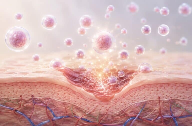
Healthy leg veins contain one-way valves. Every time your calf muscle contracts, it squeezes blood upward, and the valves snap shut behind it so that blood cannot fall back down. When those valves are damaged, weakened, or blocked, blood flows backwards and pools in the lower leg. This is called venous reflux, and the sustained high pressure it creates is called venous hypertension.
Over years, that pressure damages the smallest blood vessels in the skin. Fluid, proteins, and red blood cells leak into the surrounding tissue. The skin becomes inflamed, stiff, and poorly oxygenated. Eventually the tissue breaks down, sometimes after a minor knock, sometimes after scratching an itchy patch, and sometimes with no trigger at all.
This is why the ulcer keeps reopening even after it heals. The scratch was never the cause. The pressure was.
Where venous ulcers appear
Venous leg ulcers almost always occur in the gaiter area, the band of skin between the ankle and mid-calf, and most often on the inner side of the leg, just above the ankle bone. Location is one of the strongest clues clinicians use.
What a venous leg ulcer looks and feels like
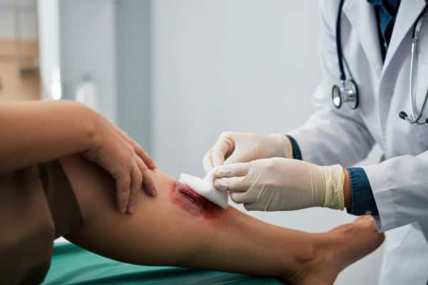
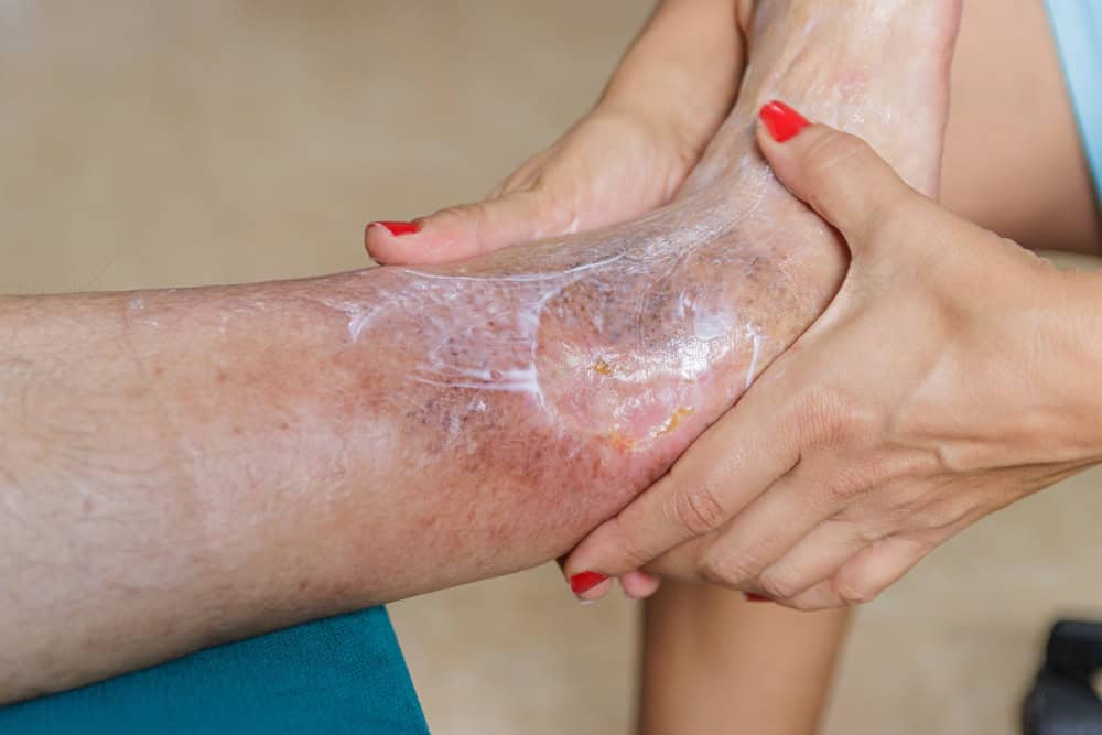
Venous ulcers have a recognisable appearance that distinguishes them from other leg wounds.
The wound itself:
- Shallow, with irregular or sloping edges rather than a clean punched-out shape
- A red or pink base, often with yellow slough (soft, stringy dead tissue) across parts of it
- Moderate to heavy fluid leakage, so dressings and socks become soaked
- An ache or heaviness rather than sharp pain, though pain varies widely
- Sometimes an unpleasant odour, particularly if infection is developing
The skin around it, often more telling than the wound:
- Hemosiderin staining: brown or rust-coloured pigmentation from iron left behind by leaked red blood cells
- Lipodermatosclerosis: hardened, tight, woody skin that narrows the lower leg, giving it an upside-down champagne bottle shape
- Venous eczema: dry, itchy, flaking, red skin that patients frequently scratch, a common route to a first ulcer
- Atrophie blanche: small smooth white patches with visible tiny vessels, marking previous damage
- Oedema: swelling that is worse by evening and better after a night lying flat
- Varicose veins: visible bulging veins, though plenty of people with venous ulcers have no obvious varicose veins at all
If you have brown staining and hard skin around your ankles but no open wound yet, that is the stage at which assessment and compression are most likely to help, and it is worth acting on.
Venous, arterial, or diabetic? Why the distinction matters
Not every leg ulcer is venous, and getting this wrong is dangerous. Firm compression applied to a leg with poor arterial supply can cause tissue death. This is the single most important reason a leg ulcer needs proper assessment before anyone wraps it.
| Venous ulcer | Arterial ulcer | Diabetic foot ulcer | |
|---|---|---|---|
| Typical location | Inner lower leg, above the ankle | Toes, heel, outer ankle, bony points | Pressure points on the sole, under the big toe or ball of foot |
| Wound edges | Irregular, shallow, sloping | Well-defined, punched out, deep | Callused rim around the wound |
| Wound base | Red or pink with yellow slough | Pale, grey, dry, may be black | Variable, often deeper than it looks |
| Fluid leakage | Moderate to heavy | Minimal | Variable |
| Pain pattern | Aching, relieved by elevating the leg | Severe, worse when elevated, better hanging down | Often painless due to nerve damage |
| Surrounding skin | Brown staining, hardened, swollen, eczema | Thin, shiny, hairless, cool, pale | Callus, dry skin, possible deformity |
| Pulses in the foot | Usually present | Weak or absent | May be present or absent |
| Cornerstone of treatment | Compression | Restore arterial blood flow | Pressure offloading |
Important: feeling a pulse does not rule out arterial disease. Around one in five people with a venous ulcer also has reduced arterial flow, and swelling makes pulses hard to feel reliably. Only an ABPI measurement can determine whether compression is safe.
Some patients have mixed disease, meaning venous insufficiency plus reduced arterial flow. These legs can often still be compressed, but at reduced pressure and under closer supervision. That decision belongs to a clinician who has measured the circulation, not to a dressing protocol.
Diabetes complicates the picture further, since neuropathy can mask pain that would otherwise warn you something is wrong. If you have diabetes and a foot or leg wound, read our guidance on diabetic wound care and diabetic foot ulcers.
Who develops venous leg ulcers
Risk accumulates over decades. Common contributing factors include:
- Previous deep vein thrombosis (DVT), which damages deep vein valves, one of the strongest single risk factors
- Varicose veins and chronic venous insufficiency
- Family history of venous disease
- Age, as valve function declines
- Obesity, which raises pressure in the abdominal and leg veins
- Prolonged standing or sitting, common in nursing, retail, hospitality, teaching, and long-haul travel
- Reduced calf muscle pump function from limited mobility, stiff ankles, paralysis, or a sedentary routine
- Previous leg fracture, surgery, or significant trauma
- Pregnancy, particularly multiple pregnancies
- Smoking, which impairs tissue repair broadly
The calf muscle pump deserves particular attention. Your calf is functionally a second heart for the lower leg. When ankle movement is restricted, through pain, arthritis, or long periods of immobility, that pump weakens, venous pressure climbs, and ulcer risk rises. This is also why walking is part of treatment rather than something to avoid.
How venous leg ulcers are diagnosed
A competent assessment goes well beyond looking at the wound.
1. History and examination. Duration of the wound, previous ulcers, DVT history, pain pattern, mobility, occupation, diabetes, and current medications. The clinician examines the skin changes described above and feels for pulses in the foot.
2. Ankle-brachial pressure index (ABPI). Blood pressure is measured at the ankle and at the arm, and the ratio is calculated. This screens for arterial disease and determines whether compression is safe. An ABPI at or below roughly 0.8 indicates significant arterial involvement, and full-strength compression should not be applied without specialist input. Compression is not used at all when arterial disease is severe (ABPI below about 0.5), or in the presence of untreated deep vein thrombosis, spreading skin infection, or uncontrolled heart failure. No leg ulcer should receive firm compression until arterial supply has been assessed.
3. Duplex ultrasound. A painless scan that maps which veins are refluxing, how severe the reflux is, and whether there is any obstruction or old clot. This is what determines whether the underlying vein problem is correctable, and it is the step most often skipped in general wound care.
4. Wound assessment and measurement. Length, width, depth, tissue type, and exudate level, recorded with photographs at each visit. Measurement matters because it converts “it looks a bit better” into data. A wound that has not reduced in area by a meaningful margin over four weeks is not responding, and that should trigger a change in plan.
5. Blood tests and swabs where indicated. To check for diabetes, anaemia, nutritional deficiency, or infection. Note that all chronic wounds carry bacteria, so a positive swab alone is not a reason for antibiotics. Clinical signs of infection are.
6. Biopsy in selected cases. An ulcer that has not healed after several months of correct treatment, or that has unusual rolled edges or excess tissue growth, may be biopsied to exclude a skin cancer masquerading as an ulcer. This is uncommon, but it is the reason non-healing ulcers deserve fresh eyes rather than another dressing change.
Treatment: what actually works
Compression therapy, the foundation
Compression is the treatment with the strongest evidence, and nothing else substitutes for it. Graduated compression counteracts venous hypertension, reduces swelling, and restores an environment where healing can occur.
In a Cochrane review of randomised trials, ulcers treated with compression healed substantially faster than those without it (pooled hazard ratio 2.17, 95% CI 1.52 to 3.10), and were about 1.8 times as likely to be fully healed within 12 months (risk ratio 1.77, 95% CI 1.41 to 2.21). The review rated this moderate-certainty evidence. That is a large effect for a non-surgical intervention.
Practical realities patients should know:
- Effective compression usually means multi-layer bandaging or medical-grade compression hosiery, not ordinary support socks from a pharmacy.
- It must be applied by someone trained in the technique. Poorly applied compression is uncomfortable and can be harmful.
- The first few days can feel tight and strange. Persistent pain, numbness, or colour change in the toes is not normal. Contact your care team the same day, and they will advise whether the bandaging needs to come off.
- Adherence is the deciding factor. Compression that is removed on hot days, on weekends, or whenever it is inconvenient does not work. In a tropical climate, this is a real challenge, and it is worth discussing lighter or more breathable systems with your clinician rather than quietly stopping.
Wound bed preparation
The wound surface must be able to support new tissue.
- Debridement removes dead tissue and slough. It may be done with dressings that soften debris, sharp instruments, or specialised techniques, depending on the wound and the patient.
- Biofilm disruption matters in long-standing ulcers. Biofilm is a protective bacterial layer that resists both antibiotics and the body’s immune response, and it needs to be physically disrupted rather than treated with antibiotics alone.
- Moisture balance. Too dry and cells cannot migrate, too wet and the surrounding skin macerates and breaks down. Dressing choice is a means to this end, not the treatment itself.
- Skin care around the wound. Emollients for venous eczema, barrier protection against exudate, and treating the itch so scratching does not create the next ulcer.
Treating the underlying vein disease
This is where standard leg ulcer care most often falls short, and where the evidence has shifted meaningfully.
The EVRA trial, published in the New England Journal of Medicine in 2018, randomised 450 patients with venous leg ulcers to either early endovenous ablation of superficial venous reflux (within two weeks) plus compression, or compression alone with the vein procedure deferred. Results in the early-treatment group:
- Median time to healing 56 days versus 82 days
- Healing at 24 weeks 85.6% versus 76.3%
- Median ulcer-free time in the first year 306 days versus 278 days
In other words, treating the faulty veins early, rather than waiting for the ulcer to close first, healed ulcers roughly a month sooner and gave patients more ulcer-free time. The most common complications of the ablation procedure were pain and deep vein thrombosis, with DVT reported more often in the early-treatment group. That trade-off is worth discussing with your clinician.
Longer term, the earlier ESCHAR trial found that adding surgical correction of superficial venous reflux to compression reduced four-year ulcer recurrence from 56% to 31%.
The practical implication is straightforward. If you have a venous leg ulcer, ask whether you have had a duplex scan and whether your superficial venous reflux is treatable. Modern options are typically minimally invasive and performed under local anaesthetic.
Managing infection appropriately
Chronic wounds are always colonised with bacteria. Antibiotics are indicated when there are clinical signs of infection, such as spreading redness, increasing pain, warmth, fever, sudden increase in exudate or odour, or rapid deterioration, not on the basis of a routine swab. Overuse drives resistance without accelerating healing.
Supporting factors that genuinely matter
- Elevation: legs raised above heart level for periods during the day, and the foot of the bed slightly raised at night.
- Walking and ankle exercises: movement drives the calf pump. Even seated ankle circles help when walking is limited.
- Nutrition: adequate protein, and correcting deficiencies in iron, vitamin C, or zinc where present. Healing has metabolic requirements.
- Diabetes and blood pressure control.
- Stopping smoking.
- Weight management, which reduces venous pressure directly.
What to expect: realistic timelines
Patients are rarely given honest numbers, which is why so many lose confidence in treatment that is actually working.
| Stage | What to expect |
|---|---|
| Weeks 1 to 2 | Swelling reduces, exudate often decreases, wound may look unchanged in size |
| Week 4 | A meaningful reduction in wound area should be visible. This is the key checkpoint |
| Week 12 | Around half of ulcers are healed in well-run services |
| Week 24 | Around 76% are healed (specialist nurse-led service, 1,324 legs) |
| Beyond 24 weeks | The ulcer is classed as hard-to-heal and warrants reassessment of the diagnosis and plan |
Healing rates differ substantially between services and patient groups, so treat these as reference points rather than promises.
The four-week checkpoint is worth raising with your care team. Less than around 30% reduction in wound area after a month predicts poor healing, and it should prompt a review rather than more of the same. The possible explanations: the arterial supply may be inadequate, there may be undiagnosed reflux or obstruction, infection or biofilm may be unresolved, compression may not be achieving therapeutic pressure, or the diagnosis itself may be wrong.
Continuing the same dressing regimen for six months without reassessment is the most common failure in leg ulcer care.
When a venous leg ulcer will not heal
Ulcers that persist despite correct compression, debridement, and vein assessment are described as hard-to-heal or recalcitrant. At this point the question changes from “which dressing?” to “what is blocking healing?”
Systematic reassessment should cover:
- Arterial supply, with a repeat ABPI or arterial imaging, particularly in older patients or those with diabetes
- Venous anatomy, with a repeat duplex, including assessment for deep vein obstruction, which is often missed
- Compression effectiveness, meaning whether therapeutic pressure is genuinely being achieved and maintained
- Infection and biofilm
- Nutrition, anaemia, and glycaemic control
- Medications that impair healing, such as long-term corticosteroids
- Alternative diagnoses, including vasculitis, pyoderma gangrenosum, malignancy, or infections that mimic venous ulcers
Adjunctive therapies
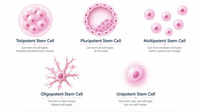
For ulcers that fail to respond to well-delivered standard care, a number of additional treatments may be considered alongside, never instead of, compression and wound bed preparation. Cleveland Clinic lists these as options for severe or non-responding venous ulcers: growth factor therapy, hyperbaric oxygen therapy, lymphoedema therapy, skin grafting, stem cell therapy, and treatment of the underlying venous disease.
Two points of honesty are important here. First, the evidence base for regenerative and biologic adjuncts is still developing, and the quality of that evidence varies considerably by therapy. Second, none of them compensates for inadequate compression or an untreated arterial problem. Any clinic offering an advanced therapy for a leg ulcer should be able to show you their assessment findings, explain why standard care has been judged insufficient, and set measurable goals before starting.
If you are being offered an adjunctive therapy, reasonable questions are: What is my ABPI? Have I had a duplex scan, and what did it show? What are we measuring, and at what interval? What would tell us this is not working?
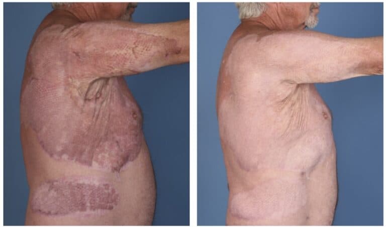
At Vegaderma, venous leg ulcers are managed within our chronic wound and skin repair pathway, which combines assessment, staged wound preparation, and compression strategies, with regenerative support considered only where clinically indicated. Documented wound measurements and serial photography are part of every case, and our published case studies show what that documentation looks like in practice.
Preventing recurrence, the part most patients are not told about
Healing the ulcer is only half the task. The venous hypertension that caused it does not disappear when the skin closes.
Reported recurrence rates vary widely between studies and services. One long-term Australian cohort found roughly 22% recurrence within three months, 57% within twelve months, and 78% within three years, with a median time to recurrence of around 42 weeks. A UK specialist nurse-led service reported far lower one-year recurrence, at around 17%. The gap between those figures is largely about follow-up quality and compression adherence, which is the point.
Recurrence falls substantially with the right follow-through:
1. Lifelong compression hosiery. This is the highest-value habit after healing. Stockings should be professionally measured, replaced roughly every three to six months as elasticity degrades, and worn daily. Application aids exist and are worth asking for if hand strength or reach is limited.
2. Correcting the underlying reflux. The ESCHAR trial showed four-year recurrence dropping from 56% to 31% when superficial reflux was surgically corrected alongside compression. Note that ESCHAR used open surgery rather than the endovenous techniques common today, and it reduced recurrence without speeding healing. If your reflux has not been assessed and treated, that is the conversation to have.
3. Daily skin care. The skin in the gaiter area stays fragile after healing. Regular emollients, prompt treatment of venous eczema, and protecting the leg from knocks all reduce the chance of a new breakdown.
4. Keeping the calf pump working. Daily walking, ankle mobility exercises, and avoiding long unbroken periods of standing or sitting.
5. Elevation and weight management as ongoing habits rather than temporary measures.
6. Acting immediately on early signs. New redness, itching, a small blister, or a graze in an area of previous ulceration should be reviewed quickly. A recurrent ulcer caught in the first week is a very different problem from one caught at six weeks.
When to seek urgent medical attention
Arrange same-day review if you notice:
- Rapidly spreading redness, warmth, or swelling around the wound
- Fever, chills, or feeling generally unwell
- A sudden increase in pain, particularly if the pain becomes severe
- Sudden increase in fluid, pus, or a marked change in odour
- Black or grey tissue appearing in or around the wound
- Toes that become pale, blue, cold, or numb after compression is applied
- Rapid enlargement of the ulcer
The bottom line
Venous leg ulcers are common, slow to heal, and prone to return, but they are not a condition you simply live with. The path that works is consistent: confirm the diagnosis, check the arterial circulation, apply proper compression, prepare the wound bed, scan the veins, treat correctable reflux early, and measure progress at four-week intervals rather than hoping.
If your ulcer has been open for more than six weeks, or has not measurably improved in the last four, it is worth asking for the plan to be reviewed.
Vegaderma’s wound care team assesses chronic and non-healing wounds, including venous leg ulcers, within a structured chronic wound and skin repair pathway led by our physicians. If you would like your wound reviewed, contact our clinical team.
Medical Disclaimer: Treatment outcomes vary between individuals. The information on this page is for educational purposes only and should not be interpreted as medical advice or a guarantee of results. A physician consultation is required to determine suitability for any treatment..
Frequently Asked Questions
-
How long does a venous leg ulcer take to heal?
Most venous leg ulcers take weeks to months. In well-run services, around half are healed by 12 weeks and around 76% by 24 weeks, though rates vary between services and patient groups. An ulcer that has not measurably shrunk after four weeks of correct compression should prompt reassessment rather than continued observation.
-
Will a venous leg ulcer heal on its own?
It is very unlikely. The underlying venous hypertension does not resolve without treatment, and untreated ulcers tend to enlarge and become infected. Prompt compression and vein assessment give the best chance of closure.
-
Is a venous leg ulcer dangerous?
It can become so. Complications include cellulitis, deeper soft tissue and bone infection, bloodstream infection, and in severe neglected cases tissue loss requiring surgery or amputation. Ulcers also cause significant pain, sleep disruption, reduced mobility, social isolation, and depression. Timely treatment substantially reduces these risks.
-
What is the difference between a venous ulcer and an arterial ulcer?
Venous ulcers sit above the inner ankle, are shallow with irregular edges, leak heavily, and are surrounded by brown-stained swollen skin, and pain typically eases when the leg is raised. Arterial ulcers occur on the toes, heel, or bony points, look deep and punched out, leak little, and are extremely painful, often worse when the leg is elevated. The treatments are different and, in the case of compression, applying the wrong one can cause harm.
-
Can I use compression stockings without seeing a doctor first?
No. Compression must not be applied until arterial circulation has been assessed, usually with an ankle-brachial pressure index. In a leg with significant arterial disease, compression can restrict blood flow further and cause tissue damage.
-
Why does my venous leg ulcer keep coming back?
Because the underlying vein problem is still there. Reported one-year recurrence ranges from around 17% in well-supported specialist services to 57% in longer-term community cohorts. The two measures that reduce it most are wearing prescribed compression hosiery long term and correcting treatable venous reflux, which cut four-year recurrence from 56% to 31% in the ESCHAR trial, where the reflux was corrected surgically.
-
Should the faulty veins be treated before or after the ulcer heals?
Current evidence favours early treatment. In the EVRA trial, treating superficial venous reflux within two weeks, rather than waiting for the ulcer to close, shortened median healing time from 82 days to 56 days and increased 24-week healing from 76.3% to 85.6%.
-
Can I shower or bathe with a venous leg ulcer?
Usually yes, with the wound and bandaging protected as your care team advises. Prolonged soaking and swimming with an open wound are generally avoided. Ask for specific guidance, as it depends on your dressing system.
-
Does walking make a venous leg ulcer worse?
No, walking generally helps. Calf muscle contraction pumps blood out of the leg and reduces venous pressure. Prolonged standing still is the problem, not walking. Combine regular walking with periods of leg elevation.
-
Question
Answer
-
Does diet affect venous ulcer healing?
Indirectly but meaningfully. Healing requires adequate protein and micronutrients, and deficiencies in protein, vitamin C, zinc, or iron slow repair. Good glycaemic control matters for anyone with diabetes. Diet supports healing, but it does not substitute for compression.
References
- Robles-Tenorio A, Lev-Tov H, Ocampo-Candiani J. Venous Leg Ulcer. StatPearls. https://www.ncbi.nlm.nih.gov/books/NBK567802/
- Cleveland Clinic. Venous Ulcer: Symptoms, Causes, Treatment & Prevention. https://my.clevelandclinic.org/health/diseases/23165-venous-ulcer
- Shi C, Dumville JC, Cullum N, et al. Compression bandages or stockings versus no compression for treating venous leg ulcers. Cochrane Database of Systematic Reviews, 2021. https://www.cochranelibrary.com/cdsr/doi/10.1002/14651858.CD013397.pub2/full
- Gohel MS, Heatley F, Liu X, et al. A Randomized Trial of Early Endovenous Ablation in Venous Ulceration (EVRA). N Engl J Med. 2018;378(22):2105-2114. https://pubmed.ncbi.nlm.nih.gov/29688123/
- Gohel MS, Barwell JR, Taylor M, et al. Long term results of compression therapy alone versus compression plus surgery in chronic venous ulceration (ESCHAR): randomised controlled trial. BMJ. 2007. https://pubmed.ncbi.nlm.nih.gov/17545185/
- Gohel MS, Taylor M, Earnshaw JJ, et al. Risk factors for delayed healing and recurrence of chronic venous leg ulcers, an analysis of 1324 legs. Eur J Vasc Endovasc Surg. 2005. https://pubmed.ncbi.nlm.nih.gov/15570275/
- Finlayson K, Wu ML, Edwards HE. Identifying risk factors and protective factors for venous leg ulcer recurrence. Int J Nurs Stud. 2015. https://pubmed.ncbi.nlm.nih.gov/25801312/
- Union Internationale de Phlebologie consensus on venous leg ulcer management, 2024. https://pmc.ncbi.nlm.nih.gov/articles/PMC11380361/
- Alavi A, Sibbald RG, Phillips TJ, et al. Venous Leg Ulcers: Advanced Therapies and New Technologies. https://www.ncbi.nlm.nih.gov/pmc/articles/PMC8615583/
- He S, et al. Prevention strategies for the recurrence of venous leg ulcers: A scoping review. International Wound Journal, 2024. https://onlinelibrary.wiley.com/doi/full/10.1111/iwj.14759
- Vasudevan B. Venous Leg Ulcers: Pathophysiology and Classification. Indian Dermatol Online J. 2014;5(3):366-370. https://www.ncbi.nlm.nih.gov/pmc/articles/PMC4144244/

The Vega Derma Medical Team consists of board-certified dermatologists, specialized trichologists, and clinical researchers based in Bangkok, Thailand. Our team specializes in evidence-based regenerative dermatology, complex tissue repair, and advanced hair restoration (including precision FUE and FSE). Every protocol and article we publish is medically reviewed to ensure it meets international clinical standards, providing safe, science-backed guidance for our local and international patients.