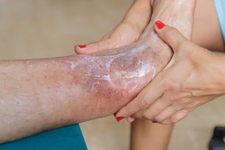

A chronic ulcer is not a wound that is healing slowly. It is a wound that has stopped healing altogether. That distinction matters more than almost anything else in this article, because it changes what treatment has to accomplish.
Effective chronic ulcer treatment has to do two jobs at the same time. First, fix the reason the wound stalled, whether that is poor blood flow, unrelieved pressure, hidden infection, or runaway inflammation. Second, rebuild the biology that closes it. Dressings on their own rarely manage either.
So if you have been changing dressings for months and the wound looks much the same as it did in week four, that is not bad luck. It is a clinical signal, and it usually means the plan needs to change rather than continue.
What follows is a plain explanation of what chronic ulcers are, why they behave differently from ordinary wounds, what good care looks like, and where advanced options such as growth factor and stem cell therapy genuinely fit. It is written for patients and families weighing up their choices, including anyone considering treatment for chronic wounds in Thailand.
What Is a Chronic Ulcer?
A chronic ulcer is an open wound that has failed to move through the normal stages of repair in the time you would expect. Most wound centres work to a simple rule of thumb:
If a wound has not healed, or has not meaningfully shrunk, after four to six weeks of appropriate care, treat it as chronic.
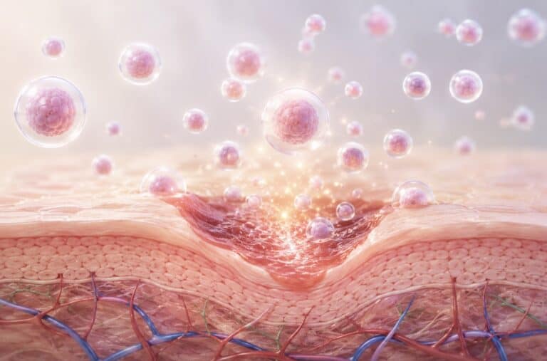
Healthy wounds pass through four phases: haemostasis, inflammation, proliferation, and remodelling. A chronic ulcer gets stuck in the inflammatory phase and stays there. The wound bed fills with destructive enzymes called matrix metalloproteinases (MMPs), which break down the same collagen scaffold that new tissue depends on. Growth factor signalling drops off. Bacteria organise themselves into protective biofilms. Between them, these changes create an environment that is actively hostile to healing.
This is the key idea behind everything else on this page. A chronic ulcer is a biological problem sitting underneath a visible wound, which is why treatment aimed only at the surface tends to plateau.
Chronic wounds are also more common than most people realise. A 2024 systematic review and meta-analysis put global chronic wound prevalence at roughly 1.67 per 1,000 people, with mixed-aetiology chronic wounds at 2.21 per 1,000. That represents tens of millions of patients and a disproportionate share of healthcare spending worldwide.
The Four Main Types of Chronic Ulcers
Chronic ulcer treatment is only ever as good as the diagnosis behind it. Two wounds that look almost identical on the lower leg can have completely different causes, and completely different correct treatments.
| Ulcer type | Typical location | Underlying driver | Treatment cornerstone |
|---|---|---|---|
| Diabetic foot ulcer (DFU) | Plantar forefoot, toes, heel | Neuropathy plus peripheral artery disease | Offloading, debridement, glycaemic control |
| Venous leg ulcer (VLU) | Above the ankle, medial gaiter area | Venous hypertension, valve failure | Graduated compression therapy |
| Pressure injury (bedsore) | Sacrum, heels, hips | Sustained pressure and shear, immobility | Pressure redistribution, repositioning |
| Arterial or ischaemic ulcer | Toes, foot margins, shin | Critical limb ischaemia | Revascularisation assessment first |
Diabetic foot ulcers carry the highest stakes of the four. Lifetime risk among people with diabetes is estimated at 19 to 34%. Recurrence reaches around 42% within one year and 65% within five years. Five-year mortality after a DFU sits near 30%, and rises above 70% following major amputation. Foot ulceration precedes most non-traumatic lower-limb amputations in people with diabetes, which is why waiting to see whether one closes on its own is rarely the safest choice.
Venous leg ulcers are the most common leg ulcer overall, affecting roughly 1% of adults and up to 4% of people over 65. Compression therapy remains the single intervention with the strongest evidence for healing them. Patients who cannot tolerate or do not stick with compression tend to take at least twice as long to heal and face far higher recurrence rates.
If your wound falls into the diabetic category, our page on diabetic foot ulcer treatment in Thailand covers Wagner staging, offloading, and regenerative options in more detail.
Why Chronic Ulcers Don’t Heal on Their Own
Five mechanisms account for most stalled wounds. A thorough treatment plan screens for all of them rather than assuming one.
Ischaemia, or poor blood supply. Oxygen and nutrients are the fuel for repair. Peripheral artery disease starves the wound bed, and no dressing compensates for absent perfusion. Vascular assessment using ABI, toe pressures, or duplex imaging should come before any advanced therapy is considered.
Neuropathy, or loss of protective sensation. In diabetes, nerve damage means blisters, callus breakdown, and pressure points go completely unnoticed. A small injury becomes a deep ulcer before the patient feels anything at all.
Unrelieved pressure. A plantar foot ulcer exposed to normal walking is re-injured with every step. Without offloading, whatever healing happens in clinic is undone at home.
Bacterial biofilm. Biofilms are structured bacterial communities wrapped in a protective matrix that resists both antibiotics and the immune system. Systemic antibiotics alone often fail against them. Sharp debridement is what physically breaks them up.
Chronic inflammation and lost signalling. The raised MMP activity and depleted growth factors described earlier mean the wound has lost its instructions for rebuilding, even when everything else has been optimised.
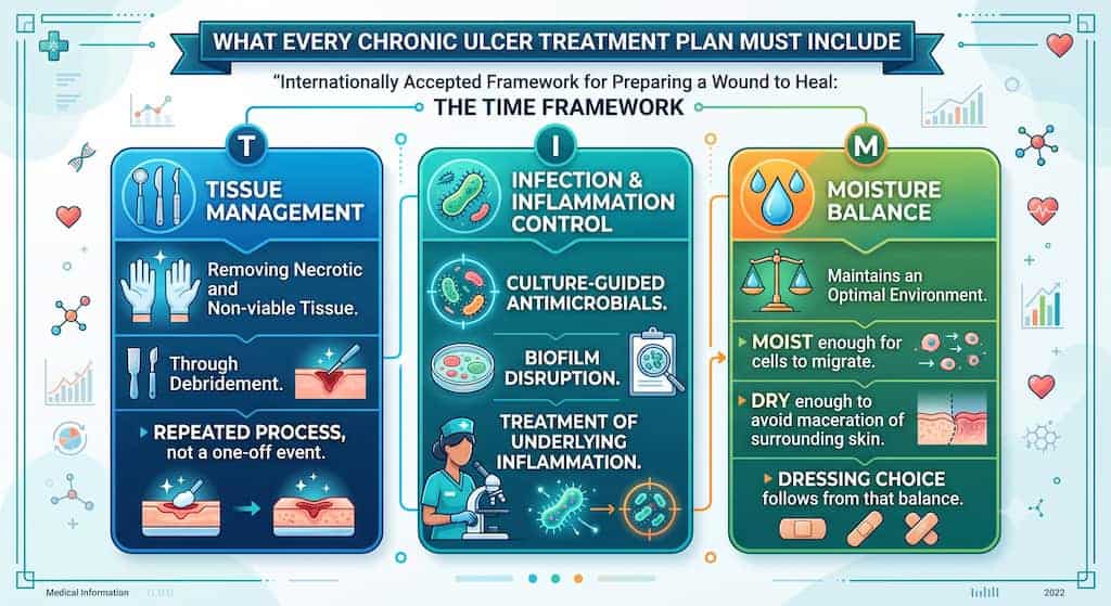
What Every Chronic Ulcer Treatment Plan Must Include
Before anything advanced enters the conversation, the fundamentals need to be in place. The internationally accepted framework for preparing a wound to heal is TIME:
- T, tissue management. Removing necrotic and non-viable tissue through debridement. This is repeated, not a one-off.
- I, infection and inflammation control. Culture-guided antimicrobials, biofilm disruption, and treatment of the underlying inflammation.
- M, moisture balance. The wound bed needs to be moist enough for cells to migrate, but dry enough to avoid maceration of the surrounding skin. Dressing choice follows from that balance.
- E, edge advancement. Watching whether the wound margin is genuinely migrating inward, which is the clearest single sign that healing has restarted.
Alongside TIME, four cause-specific measures do most of the heavy lifting:
- Offloading for diabetic foot ulcers, using total contact casting, removable walkers, or bespoke orthotics.
- Graduated compression for venous leg ulcers, which carries the highest level of evidence for VLU healing.
- Pressure redistribution and repositioning for pressure injuries.
- Systemic optimisation, covering glycaemic control, nutrition (particularly protein and micronutrients), smoking cessation, and oedema management.
Skipping these steps and jumping straight to an advanced therapy is the most common reason advanced therapies disappoint. Regenerative treatment works as a catalyst. It does not work as a substitute.
When Standard Care Isn’t Enough
If a wound has been properly prepared, offloaded, and cleared of infection, and it still has not improved after four weeks, it qualifies as hard-to-heal. At that point, escalating treatment is reasonable.
Growth Factor Therapy (VEGF and PDGF)
Two signalling proteins coordinate much of wound repair.
- VEGF (Vascular Endothelial Growth Factor) drives angiogenesis, the sprouting of new capillaries into the wound bed. It restores the oxygen and nutrient supply that ischaemic tissue is missing.
- PDGF (Platelet-Derived Growth Factor) recruits fibroblasts, the cells that produce collagen and build the new tissue scaffold.
Because the two act on different stages of the healing cascade, one on blood supply and one on tissue construction, using them together covers more of the pathway than either does alone. At Vega, this is delivered as VEGF & PDGF Ultra Enhanced Media, applied directly to a prepared wound bed during scheduled clinical visits and reviewed for response at each stage. The full pathway is set out on our chronic wound and skin repair page.
Stem Cell Treatment for Diabetic Foot Ulcers: What the Evidence Shows
Stem cell treatment for diabetic foot ulcers is one of the most actively researched areas in wound medicine, and also one of the most frequently oversold. Here is a fair reading of where the literature currently stands.
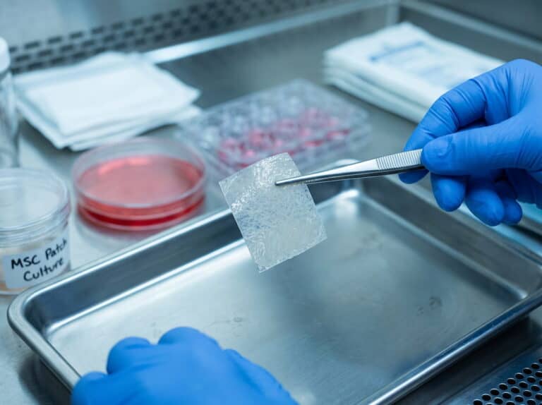
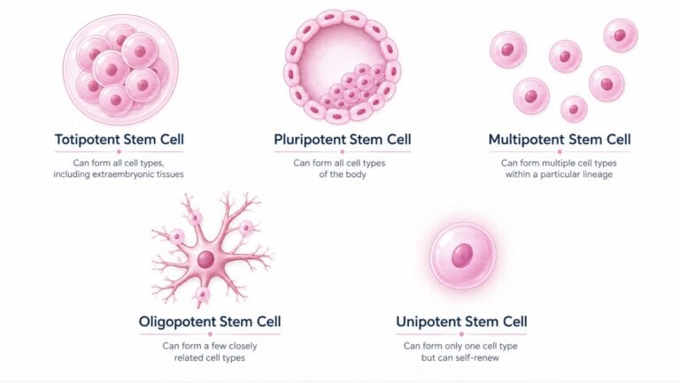
Mesenchymal stem cells (MSCs) appear to help chronic wounds mainly through paracrine signalling. Rather than becoming new skin themselves, they release growth factors and exosomes that instruct the cells already present in the wound bed. The proposed effects are promoting angiogenesis, calming excessive inflammation, and coordinating fibroblast activity.
The clinical evidence is encouraging, though still maturing:
- A meta-analysis of 28 randomised controlled trials covering 1,096 participants found autologous stem cell therapy was associated with significantly higher complete healing rates in chronic lower-extremity wounds (RR 1.67).
- A meta-analysis of 6 RCTs covering 380 patients on umbilical cord derived MSCs for diabetic foot ulcers and peripheral artery disease reported better ulcer healing (OR 2.88), along with improvements in pain, skin temperature, and ulcer area.
- Adverse events reported across these trials were generally transient, minor, and local, which supports a reasonable short-term safety profile.
The caveats deserve equal weight. Trial sizes are small. Cell sources and protocols vary widely between studies. Follow-up is often short. Regulatory status differs considerably from country to country. No credible clinic should present stem cell therapy as a guaranteed cure for a non-healing ulcer.
At Vega Derma, these regenerative principles are delivered as bio-active growth factor signalling rather than live cell implantation, within a structured, physician-supervised protocol. We state that plainly because patients researching this area deserve accuracy rather than marketing language.
Other Advanced Adjuncts
- Negative pressure wound therapy (NPWT) reduces oedema and encourages granulation in deep or heavily exuding wounds.
- Hyperbaric oxygen therapy (HBOT) is hospital-based and considered for selected ischaemic and Wagner grade 3 and above diabetic wounds.
- Cellular and tissue-based products, sometimes called skin substitutes, are bioengineered grafts for wounds with adequate perfusion.
- Platelet-rich plasma (PRP) is an autologous concentrate of the patient’s own growth factors.
Vega Derma operates as a specialist outpatient regenerative wound clinic. Cases needing inpatient surgery, revascularisation, or hyperbaric facilities are coordinated with the appropriate hospital services rather than managed in-house. That boundary is worth confirming with any clinic you are considering.
How to Tell Whether Your Treatment Is Working
You should not have to guess. There is a well-validated benchmark, often called the four-week rule:
A wound that has not reduced in area by roughly 50% after four weeks of appropriate treatment is unlikely to close on that trajectory and should be formally reassessed.
The landmark study behind this found that diabetic foot ulcers with above-median area reduction at four weeks went on to a 12-week healing rate of 58%, compared with just 9% for those below it. The dividing line fell at about 53% area reduction. Put simply, four weeks of measurement tells you far more than four months of hoping.
Making that benchmark usable requires objective tracking. At minimum, your clinic should be recording:
- Wound area in mm² at every visit, including length, width, and depth
- Serial photographic documentation taken under consistent conditions
- Tissue quality, meaning the proportion of granulation against slough or necrosis
- Exudate volume, odour, and the condition of the surrounding skin
- Edge advancement, or whether the margin is actually moving inward
If your current provider cannot show you a measured trend line, that is a fair reason to seek a second opinion. Good records also help international patients share documented progress with a referring physician or insurer back home.
Chronic Ulcer Treatment in Thailand: Why Patients Travel
Thailand has become a recognised destination for treatment for chronic wounds, for four connected reasons.
- Cost. Advanced regenerative wound care in Thailand typically costs a fraction of the equivalent in the US, UK, or Australia, with itemised quotations issued after assessment rather than open-ended billing.
- Continuity with one team. Chronic ulcer treatment is a long process. Being followed by the same specialists from assessment through to closure avoids the detail loss that happens in rotating-staff hospital clinics.
- Access. Specialist wound assessment can usually be arranged within days rather than months.
- English-speaking support, including help with scheduling, travel, and documentation for overseas patients.
One practical note for anyone planning to travel. Chronic ulcer treatment is not a single-visit procedure. Ask any clinic for an honest estimate of how many visits you will need, how far apart they fall, and how much of the care can continue at home under remote supervision, all before you book flights.
What a Structured Pathway Looks Like
| Stage | What happens |
|---|---|
| 1. Comprehensive assessment | Vascular, neurological, nutritional, and metabolic evaluation, plus wound staging (Wagner grading for diabetic foot ulcers) |
| 2. Wound bed preparation | Debridement of necrotic tissue and biofilm disruption under the TIME framework |
| 3. Infection control | Culture-guided antimicrobial strategy, with escalation to hospital care if systemic infection is present |
| 4. Regenerative support | VEGF & PDGF Ultra Enhanced Media applied to a prepared wound bed where clinically appropriate |
| 5. Mechanical management | Offloading specific to wound location, or compression for venous ulcers |
| 6. Objective monitoring | Wound measurement and serial photography at every visit, with the protocol adjusted on documented response |
| 7. Recurrence prevention | Footwear, orthotics, compression hosiery, skin care, and structured follow-up |
Candidacy matters at every stage. Regenerative support is generally suitable for chronic wounds open beyond six weeks with adequate blood supply and controlled infection. It is not appropriate as a standalone therapy for wounds requiring urgent surgery, for severe ischaemia awaiting vascular intervention, or during active uncontrolled infection. In those cases the right first step is hospital referral, with reassessment afterwards.
Preventing Recurrence After the Ulcer Closes
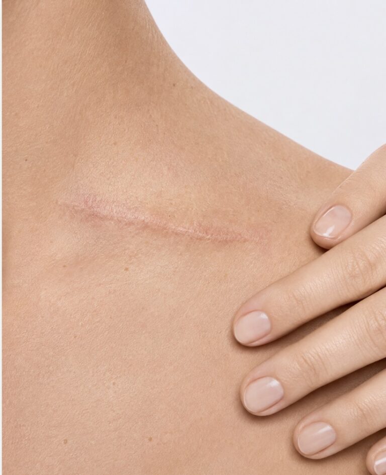
Closure is a milestone rather than an endpoint. The circulation and nerve changes that caused the first ulcer do not disappear once the skin seals over, which is why a previously ulcerated foot is formally classified as at-risk, or Wagner Grade 0.
Prevention that has evidence behind it includes:
- Daily foot inspection for blisters, redness, callus, or new pressure points
- Professional nail and callus care, never self-treated in a neuropathic foot
- Therapeutic footwear and custom orthotics to redistribute plantar pressure
- Lifelong compression hosiery after a healed venous ulcer, where the difference in recurrence rates between compression and none is substantial
- Tight glycaemic control and sustained nutritional support
- Prompt review of any new wound, however minor it looks
Frequently Asked Questions
When does a wound become a chronic ulcer?
Most wound centres classify a wound as chronic once it has not healed after roughly four to six weeks of appropriate care. A wound that stops shrinking, keeps reopening, or becomes repeatedly infected should be assessed as chronic regardless of exactly how long it has been there.
What is the best treatment for a chronic ulcer?
There is no single best treatment, because the right plan depends on why the wound stalled. The strongest results come from combining debridement, infection and biofilm control, moisture balance, cause-specific mechanical management such as offloading or compression, and, for wounds that remain stalled, regenerative support such as growth factor therapy.
Can stem cell therapy heal a non-healing ulcer?
Research suggests stem cell based approaches may support healing in selected cases by improving cellular signalling, encouraging new blood vessel formation, and reducing chronic inflammation. Meta-analyses report better healing rates than conventional care alone, though trials remain small and protocols vary. It is used alongside standard wound care, never as a replacement for debridement and infection control.
How long does chronic ulcer treatment take?
Timelines vary widely. Smaller, well-perfused ulcers may show clear improvement within a few weeks once infection is controlled and pressure is relieved. Deep or long-standing wounds can take several months, and progress depends heavily on blood sugar control, perfusion, and how consistently offloading or compression is maintained.
Is my treatment failing if the ulcer hasn’t closed yet?
Not necessarily, but it should be measurably smaller. If the wound has not reduced in area by roughly half after four weeks of appropriate treatment, the plan should be formally reassessed rather than simply continued at the same intensity.
What does chronic ulcer treatment cost in Thailand?
Cost depends on ulcer type and stage, infection status, debridement requirements, the number of visits, and whether regenerative therapy is included. Reputable clinics assess first and quote second. Vega provides an itemised estimate after clinical assessment rather than a fixed package price.
Can a chronic ulcer be treated without surgery?
Many can. Most chronic ulcers are managed through structured outpatient wound care. Surgery becomes necessary for deep infection, osteomyelitis, gangrene, or when revascularisation is needed to restore blood supply, which is exactly why vascular assessment forms part of the initial workup.
The Bottom Line
Chronic ulcer treatment works when it is diagnostic before it is therapeutic. Work out why the wound stalled, correct that cause, prepare the wound bed properly, take the mechanical load off it, and only then bring in regenerative support to restart the biology.
If your wound has stayed open beyond four to six weeks despite consistent care, that is the moment to seek specialist assessment rather than repeat the same routine for another month.
Book a wound assessment with Vega’s clinical team to have your case staged and a documented plan prepared. You can also read more about our chronic wound and skin repair programme or our diabetic foot ulcer treatment pathway.
Reviewed by Vega’s wound care specialists, who bring over 15 years of combined clinical experience in regenerative wound management. Meet our doctors.
References:
- Martinengo L, et al. Prevalence of chronic wounds in the general population: systematic review and meta-analysis of observational studies. Annals of Epidemiology. https://pubmed.ncbi.nlm.nih.gov/30497932/
- Digital Health Interventions for Chronic Wound Management: A Systematic Review and Meta-Analysis. Journal of Medical Internet Research, 2024. https://www.jmir.org/2024/1/e47904
- Armstrong DG, Boulton AJM, Bus SA. Diabetic Foot Ulcers and Their Recurrence. New England Journal of Medicine, 2017. https://www.nejm.org/doi/full/10.1056/NEJMra1615439
- Armstrong DG, Tan TW, Boulton AJM, Bus SA. Diabetic Foot Ulcers: A Review. JAMA, 2023. https://pmc.ncbi.nlm.nih.gov/articles/PMC10723802/
- Diabetic Foot Ulceration and Complications. StatPearls, NCBI Bookshelf. https://www.ncbi.nlm.nih.gov/books/NBK499887/
- Autologous Stem Cell Therapy for Chronic Lower Extremity Wounds: A Meta-Analysis of Randomized Controlled Trials. https://www.ncbi.nlm.nih.gov/pmc/articles/PMC8699089/
- Zhang H, Gu Y, Zhang K, Ouyang C. Safety and Efficacy of Umbilical Cord Mesenchymal Stem Cell Therapy for Diabetic Foot Ulcers and Peripheral Artery Disease: A Systematic Review and Meta-Analysis of Randomized Controlled Trials. 2026. https://doi.org/10.1177/15347346261426552
- Advancements in mesenchymal stem cell therapy for chronic wounds: challenges, innovations, and future directions. Frontiers in Cell and Developmental Biology. https://pmc.ncbi.nlm.nih.gov/articles/PMC12832901/
- Efficacy and safety of stem cell therapy in patients with chronic lower extremity ulcers: an umbrella review of systematic reviews and meta-analyses. Stem Cell Research & Therapy. https://link.springer.com/article/10.1186/s13287-025-04853-x
- He Y, et al. Prevention strategies for the recurrence of venous leg ulcers: A scoping review. International Wound Journal, 2024. https://onlinelibrary.wiley.com/doi/full/10.1111/iwj.14759
- Compression therapy for preventing venous leg ulcers returning. Cochrane Review. https://www.cochrane.org/evidence/CD002303_compression-therapy-preventing-venous-leg-ulcers-returning
- VenUS IV, compression hosiery compared with compression bandaging in the treatment of venous leg ulcers. NIHR Journals Library / NCBI Bookshelf. https://www.ncbi.nlm.nih.gov/books/NBK262504/
- Sheehan P, Jones P, Caselli A, Giurini JM, Veves A. Percent Change in Wound Area of Diabetic Foot Ulcers Over a 4-Week Period Is a Robust Predictor of Complete Healing in a 12-Week Prospective Trial. Diabetes Care, 2003. https://diabetesjournals.org/care/article/26/6/1879/26496/Percent-Change-in-Wound-Area-of-Diabetic-Foot

The Vega Derma Medical Team consists of board-certified dermatologists, specialized trichologists, and clinical researchers based in Bangkok, Thailand. Our team specializes in evidence-based regenerative dermatology, complex tissue repair, and advanced hair restoration (including precision FUE and FSE). Every protocol and article we publish is medically reviewed to ensure it meets international clinical standards, providing safe, science-backed guidance for our local and international patients.