
A keloid is far more than an oversized scar. It actively grows beyond the boundaries of the original wound, rarely fades on its own, and frequently returns if it is not managed properly. These traits are what separate a keloid from ordinary scarring — and why lasting removal almost always requires more than one treatment working together.
The different types of keloid scars vary widely in shape, size, and location on the body. Because treatment intensity and recurrence risk differ from one type to the next, an accurate clinical assessment is the essential first step toward a plan that produces meaningful, durable results.
Quick answer: The recognized types of keloid scars are papular, nodular, linear, flat, pedunculated, and tumoral (massive) keloids. They are grouped by how they look and how they grow, and each type responds differently to treatment.
How Keloids Differ From Other Scars
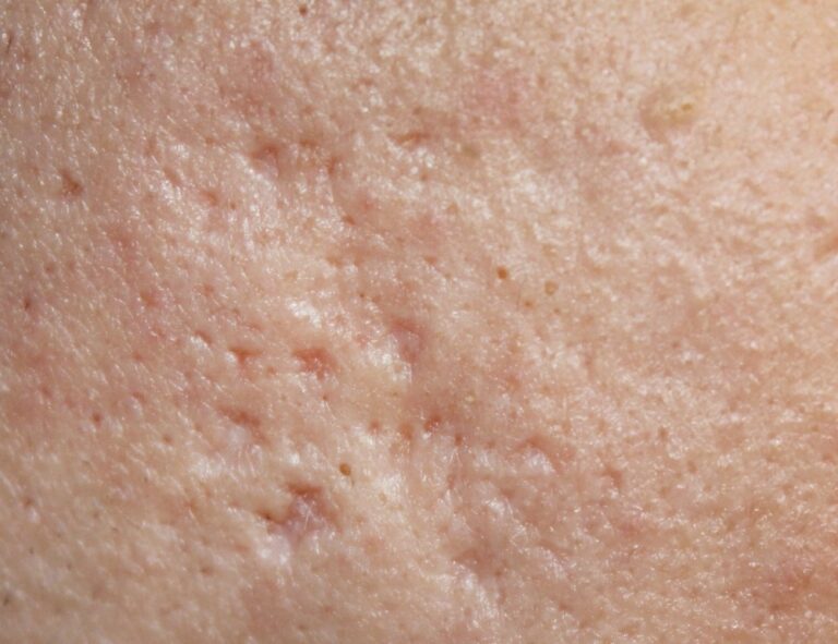
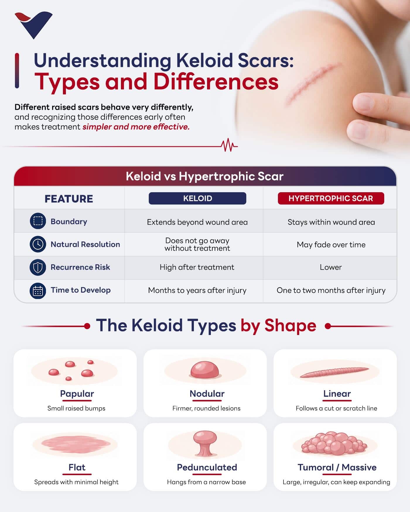
When skin heals, the body lays down collagen to close the wound. In some people that process does not switch off on time and continues past the wound edge, producing an abnormal, overgrown scar. Keloids are the most distinctive example — and the most commonly confused with hypertrophic scars, which look similar at first glance but behave very differently.
| Feature | Keloid Scar | Hypertrophic Scar |
|---|---|---|
| Growth | Extends beyond the original wound | Stays within the wound boundary |
| Onset | Months to a year (or more) after injury | Usually within 1–2 months |
| Colour | Red to purple | Pink to red |
| Natural course | Does not fade on its own | Often flattens or fades over time |
| Recurrence | High — commonly returns after treatment | Lower — generally easier to treat |
Understanding this difference matters clinically: a hypertrophic scar may resolve with conservative care, while a keloid typically persists and resists treatment unless it is addressed with a targeted, layered plan.
Types of Keloid Scars
Keloids are classified by shape and growth pattern, which directly influences how each one is treated. The main categories include:
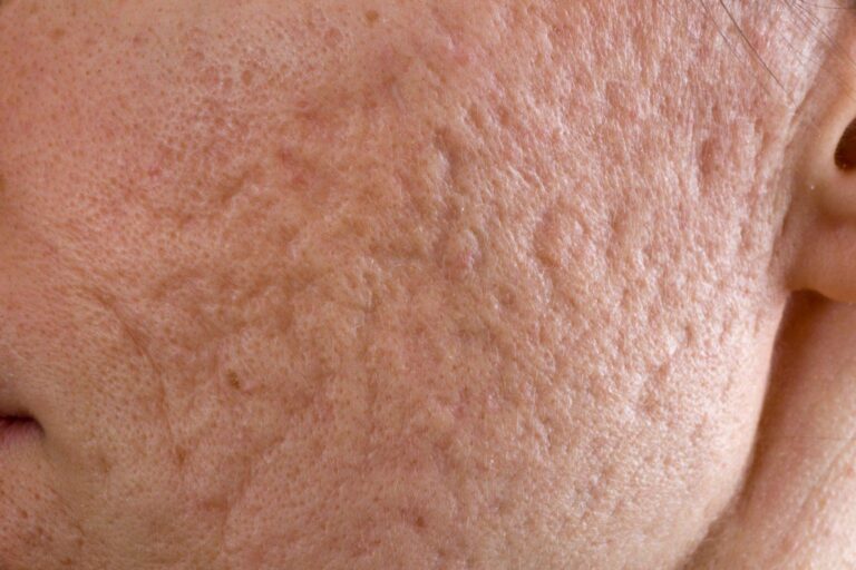
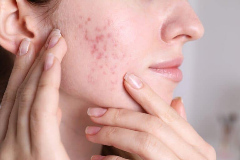
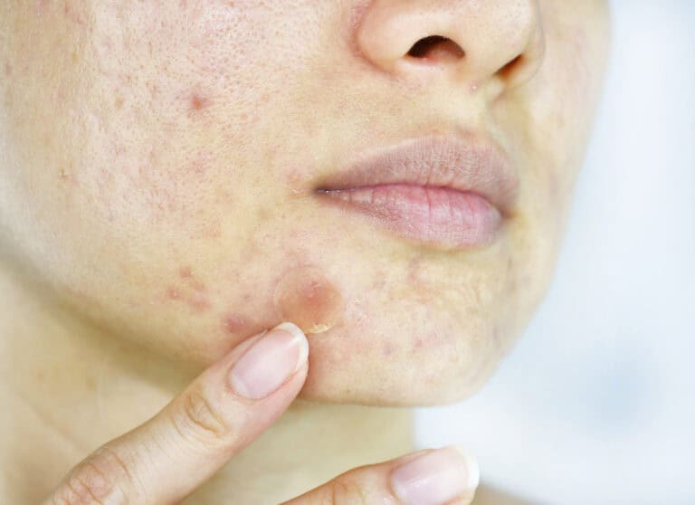
- Papular keloids: Small, raised bumps in scattered clusters, often appearing after acne or minor skin injuries.
- Nodular keloids: Firmer, rounded lesions with a defined shape, typically denser to the touch than papular forms.
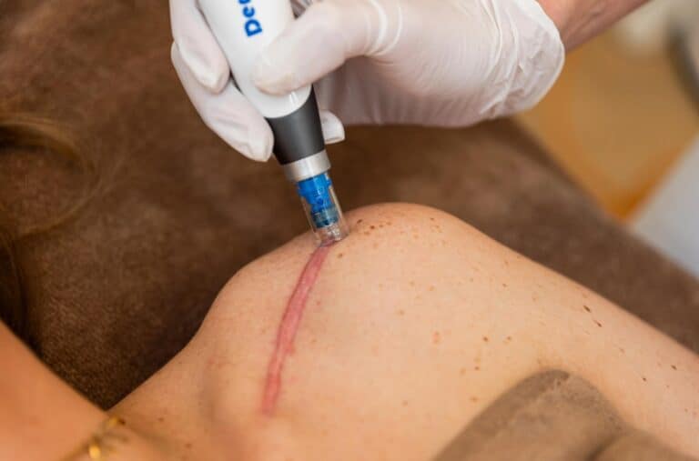
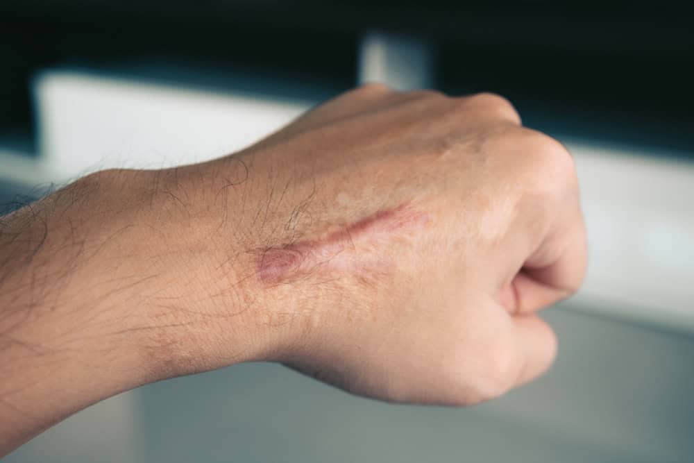
- Linear keloids: Follow a cut, scratch, or piercing track — often seen along surgical incisions in susceptible patients.
- Flat keloids: Spread across the skin surface without much height, yet still extend beyond the original wound boundary.
- Pedunculated keloids: Hang from a narrow base of tissue, most commonly on the earlobes after piercings.
- Tumoral or massive keloids: Large, irregular lesions that can keep expanding and may restrict joint movement or interfere with daily function.
Accurate classification is important because treatment intensity, expected outcomes, and recurrence risk vary substantially between these types. A small papular keloid and a massive tumoral keloid are effectively two different clinical problems.
What Causes Keloid Scars to Form?
Keloids form when the skin’s normal repair process overproduces collagen during healing. Instead of stopping once the wound has closed, fibroblasts (the cells that build scar tissue) keep working, and the scar continues to expand outward and upward.
The exact reason some people scar this way is not fully understood, but several factors are consistently linked to keloid formation:
- Genetics and family history — a strong predictor of who will develop keloids.
- Skin tone — keloids are observed more frequently in people with darker skin.
- Wound tension — injuries in high-movement, high-tension areas are more prone to keloids.
- Minor skin trauma — a piercing, pimple, insect bite, burn, or small cut can be enough to trigger one in a susceptible person.
Where Keloids Form and Who Is at Risk
The ears, chest, shoulders, upper back, and jawline are the most common sites for keloid formation. Skin in these areas is frequently under mechanical tension, which encourages continued collagen activity and makes keloids here more likely to recur after treatment.
Risk factors that increase the likelihood of developing a keloid include:
- Darker skin tones, where keloid scarring is more frequently observed clinically.
- Family history of keloids, which carries a strong genetic component.
- Skin injuries in high-tension areas of the body.
- Minor triggers such as a piercing, a pimple, or a small cut in susceptible individuals.
Common Symptoms and Physical Effects of Keloids
Beyond the visible scar, keloids can cause a range of physical symptoms that affect daily comfort. Common presentations include:
- Itching, burning, or tenderness in and around the scar tissue.
- Tightness or restricted movement when the keloid forms near a joint or on highly mobile skin.
- Active or inflamed keloids that appear red, warm, and painful — reflecting ongoing excess collagen activity beneath the surface.
Keloids on visible areas such as the face, ears, or neck also carry a cosmetic and psychological impact that is worth discussing with a physician as part of any treatment plan.
Medical Treatment for Keloids
Most keloids respond best to combination care. Here is how the main evidence-based options compare.
| Treatment | How it works | Best for | Typical course | Recurrence note |
|---|---|---|---|---|
| Corticosteroid injections | Reduce collagen + inflammation | Small–medium keloids | Monthly series | Lower when the full series is completed |
| Laser therapy | Improves redness, thickness, texture | Colour/surface concerns | Multiple sessions | Often paired with injections |
| Cryotherapy | Freezes and shrinks scar tissue | Small, early keloids | 1–3+ sessions | Often combined with injection |
| Surgical excision | Removes scar bulk | Larger/pedunculated keloids | Single procedure + adjuvant | High if done alone |
| Silicone + pressure therapy | Hydration + steady pressure | Post-procedure support | Daily, months | Supports long-term control |
| Cell-based regenerative support | Aims to encourage more organised collagen | Select post-treatment cases | Per protocol | Emerging; adjunctive only |
Corticosteroid (Triamcinolone) Injections — First Line
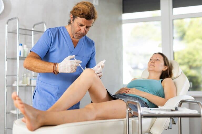
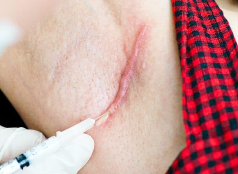
Intralesional corticosteroid injections are the standard first-line medical treatment for keloids. They soften and flatten the scar by reducing collagen production and local inflammation, and are usually given monthly until the scar improves. Possible side effects include skin thinning, pigment change at the injection site, and short-term discomfort. Injections are frequently combined with laser, cryotherapy, or — after removal — as an adjuvant.
Laser Therapy
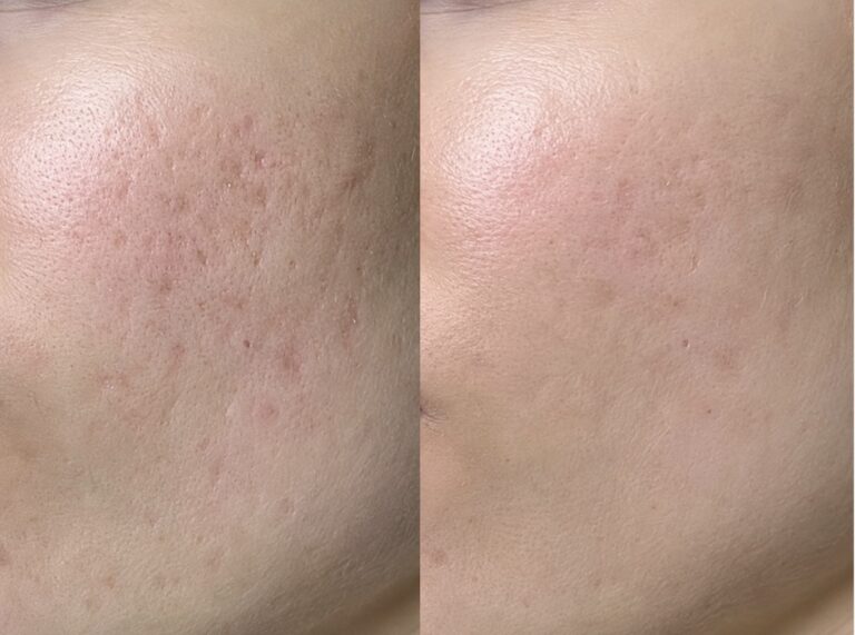
Laser treatment improves redness, thickness, and surface texture, and results are generally stronger when it is combined with steroid injections rather than used alone.
Cryotherapy
Cryotherapy freezes the keloid to shrink it and is most useful for small, early keloids, often paired with injection. It can cause temporary pigment change, particularly in darker skin — a key consideration for higher-Fitzpatrick patients.
Emerging Option: Cell-Based & Regenerative Support
Beyond the established treatments above, there is growing research interest in cell-based and regenerative approaches to scar management. The idea is that mesenchymal-cell signalling may help modulate fibroblast activity and collagen output, supporting more organised healing after primary treatment.
What’s important to understand:
- This is an evolving area of clinical research, not established standard keloid care. Current evidence is largely preclinical or early-stage human data (small pilot studies and case reports), and outcomes vary.
- It is best thought of as adjunctive — supportive of proven therapies, not a replacement for them, and not a standalone “keloid removal” method.
- Suitability must be physician-assessed for the individual scar and patient.
At Vega, cell-based regenerative support is offered only within select, staged treatment plans — after and alongside primary therapy — as part of an integrated scar and tissue remodeling program. Learn more about how this fits a personalised plan on our keloid treatment page.

Can Keloids Be Prevented?
Keloids cannot always be prevented, but people with a known history or family risk can lower the odds:
- Avoid elective skin trauma — think carefully before piercings, tattoos, or non-essential surgery on high-risk areas.
- Treat wounds early and gently — keep new wounds clean, moisturised, and protected while they heal.
- Use silicone early — starting silicone gel or sheets on a healing wound may help reduce abnormal scarring in at-risk patients.
- Flag your history to your surgeon — if you scar easily, tell any doctor before a planned procedure so they can plan preventive measures.
Why Persistent Keloids Need a Structured Clinical Approach
A keloid that keeps growing, becomes more painful, or returns after previous treatment is a clear sign that the underlying drivers have not been fully addressed. Specialist evaluation is strongly recommended for scars that:
- Restrict joint movement or interfere with daily activities.
- Appear on highly visible areas like the face, ears, or neck.
- Have already returned after one or more prior treatments.
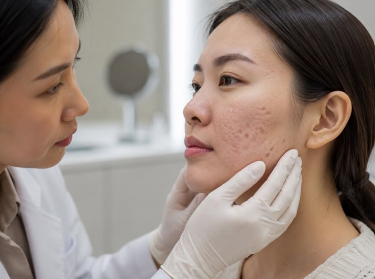
Complex keloids are best assessed by specialists who evaluate the scar type, location, and activity level together before repeating any treatment. At Vega Derma Clinic in Bangkok, the clinical team designs a staged, tailored treatment plan built around exactly what each keloid requires. Consultations are available in English for both local and international patients.
Persistent scars deserve a proper medical review. Book a consultation today to have your keloid assessed and identify the most effective course of treatment.

The Vega Derma Medical Team consists of board-certified dermatologists, specialized trichologists, and clinical researchers based in Bangkok, Thailand. Our team specializes in evidence-based regenerative dermatology, complex tissue repair, and advanced hair restoration (including precision FUE and FSE). Every protocol and article we publish is medically reviewed to ensure it meets international clinical standards, providing safe, science-backed guidance for our local and international patients.