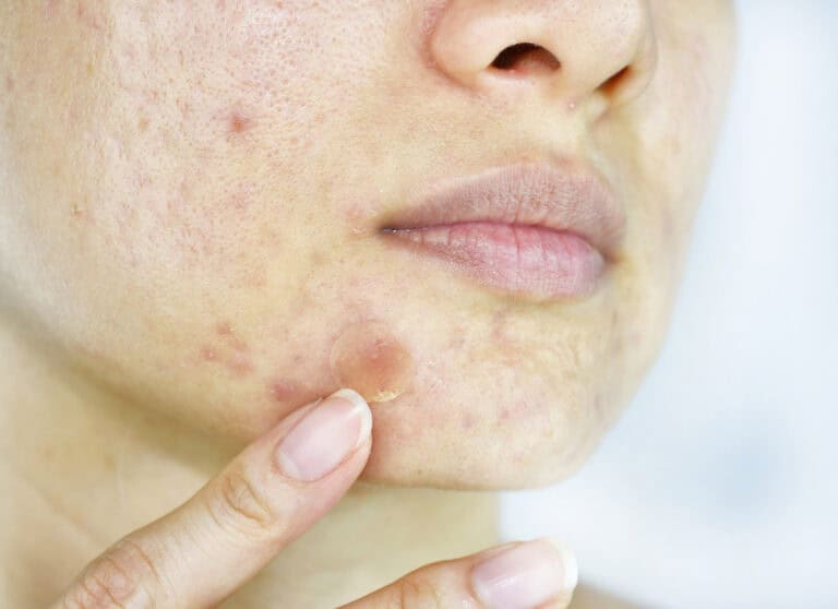

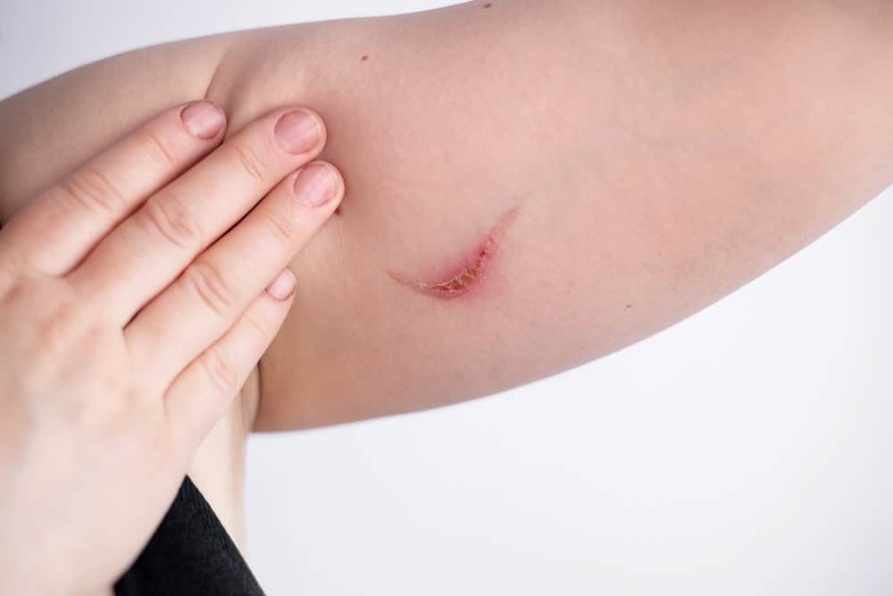
Burns are among the most common skin injuries worldwide — the World Health Organization estimates that burns cause roughly 180,000 deaths each year, with the vast majority occurring in low- and middle-income countries. Yet most burns people encounter day to day are mild and heal well when handled correctly.
Understanding the differences between first-, second-, and third-degree burns may help patients and caregivers respond appropriately when an injury happens and recognize when professional care is needed.
The single most useful thing to know about any burn is its degree, because depth determines almost everything that follows: how it heals, what risks it carries, how much it hurts, and what treatment is appropriate. This guide explains the differences between 1st, 2nd, and 3rd degree burns so you can respond appropriately and recognize when professional care is needed.
How Burn Degrees are Classified
Burns are classified by depth, meaning how deeply the injury extends into the skin and the tissues beneath. The skin is a layered organ, and the burn degree corresponds directly to how many of those layers have been affected. The deeper the burn, the more biological repair the body must perform, and the higher the risk of infection, scarring, fluid loss, and longer-term complications.
Layers of the Skin Affected by Burns
Understanding the three primary tissue layers makes burn classification easier to follow.
- Epidermis: the outermost protective layer, responsible for barrier function and the visible appearance of skin. It contains no blood vessels of its own.
- Dermis: the middle layer that contains blood vessels, nerves, hair follicles, sweat glands, and most of the structures responsible for skin regeneration.
- Hypodermis and deeper tissues: subcutaneous fat, muscle, and (in the most severe cases) bone, which have very limited ability to repair without surgery.
Mapped to depth:
- A first-degree burn affects only the epidermis.
- A second-degree burn extends into the dermis.
- A third-degree burn passes through both, and deeper injuries can reach underlying tissue.
This depth-based framework — the classical three-tier classification — remains the most widely used system in clinical assessment.
First-Degree Burns (Superficial Burns)
First-degree burns are the mildest category and the type most people meet in everyday life. The injury sits entirely in the epidermis, so the deeper structures responsible for sensation and regeneration stay intact. Healing is usually straightforward — but recognizing a first-degree burn correctly matters, because not every superficial-looking burn is as mild as it appears.
Symptoms
- Redness — the most visible sign, from increased blood flow to the area.
- Mild swelling, usually subtle and resolving within a day or two.
- Mild pain or tenderness, generally controlled with over-the-counter pain relief.
- Dry skin with no blisters. If blisters appear, the burn has reached the dermis and should be reassessed as second-degree.
Severity at a glance
- Affects only the epidermis.
- The mildest burn type; usually managed at home.
- Heals within about 3–6 days, typically without scarring.
Common causes: mild sunburn, or brief contact with a hot surface (a pan handle, iron, or hot seatbelt buckle).
Home care
- Apply cool (not cold) water or compresses to ease discomfort. Avoid ice, which can worsen tissue damage.
- Use a gentle moisturizer to support the skin barrier.
- Take OTC pain relief if needed.
- Protect the area from further sun or heat while it heals.
Second-Degree Burns (Partial-Thickness Burns)
Second-degree burns are more serious. The injury reaches past the outer layer into the dermis, where nerves and blood vessels sit — which is why these burns are significantly more painful and need more careful wound care. Healing time and scarring risk depend on how deep into the dermis the burn extends (superficial vs. deep partial-thickness).
Symptoms
- Blisters — the most distinctive sign, formed as fluid collects between damaged layers. The intact blister roof acts as a natural protective barrier.
- Significant pain, as dermal nerve endings are exposed.
- Appearance is typically red, pink, or mottled, often moist or weeping.
- Swelling at and around the wound, sometimes beyond the visibly burned area.
Severity at a glance
- Extends into part of the dermis.
- Often needs medical assessment for large burns, sensitive locations, or vulnerable patients.
- Infection risk rises once blisters break or the wound is contaminated.
- Pigmentation changes may persist, especially in darker skin tones.
- Superficial second-degree burns often heal without major scarring; deeper ones commonly scar.
- Healing time: roughly 2 weeks for superficial, several weeks for deep partial-thickness.
Common causes: scalds from hot liquids (water, coffee, cooking liquids), blistering sunburn, brief flame contact, hot grease splashes.
Wound care
- Clean gently with cool water rather than harsh antiseptics.
- Do not deliberately break blisters — the intact roof protects against infection.
- Apply a clinician-recommended dressing and watch for infection signs (spreading redness, warmth, pus, or fever).
- Seek medical evaluation for burns that are large, or on the face, hands, feet, joints, or genitals.
- Once the wound closes, structured scar care may be appropriate for deeper burns.
Third-Degree Burns (Full-Thickness Burns)
Third-degree burns — also called full-thickness burns — are the most severe category in routine clinical classification. The injury passes completely through the epidermis and dermis, destroying not just the skin’s barrier but the structures that regenerate it.
Because the body cannot rebuild tissue at this depth on its own, full-thickness burns almost always require specialist care and frequently involve surgical reconstruction. Recognizing one quickly is critical — delayed treatment increases the risk of infection, fluid loss, and lasting functional impairment.
Symptoms
- Appearance may be white, brown, black, or charred rather than red.
- Texture is often dry, stiff, leathery, or waxy.
- Reduced sensation at the center, because the nerve endings have been destroyed — while less-damaged edges may hurt more.
- A key warning sign: a burn that feels numb in the middle but painful at the edges may be far deeper than it looks.
Severity at a glance
- Extends through the full thickness of the skin; may reach fat or deeper tissue.
- High risk of infection, fluid loss, and systemic effects, especially over larger body areas.
- The body cannot regenerate full-thickness skin unaided — specialist intervention is almost always required.
- Long-term issues include scarring, contractures (tightening that limits movement), and rehabilitation that can take months or years.
Common causes: prolonged flame exposure, electrical burns (where surface injury understates internal damage), deep chemical burns, and prolonged scalds.
Treatment
- Emergency medical care is required — home management is not appropriate.
- Skin grafting or reconstructive surgery is often needed to close the wound.
- Multidisciplinary follow-up for infection and functional recovery.
- Long-term rehabilitation for contractures and scarring near joints.
- Ongoing scar care may continue for months or years after closure.
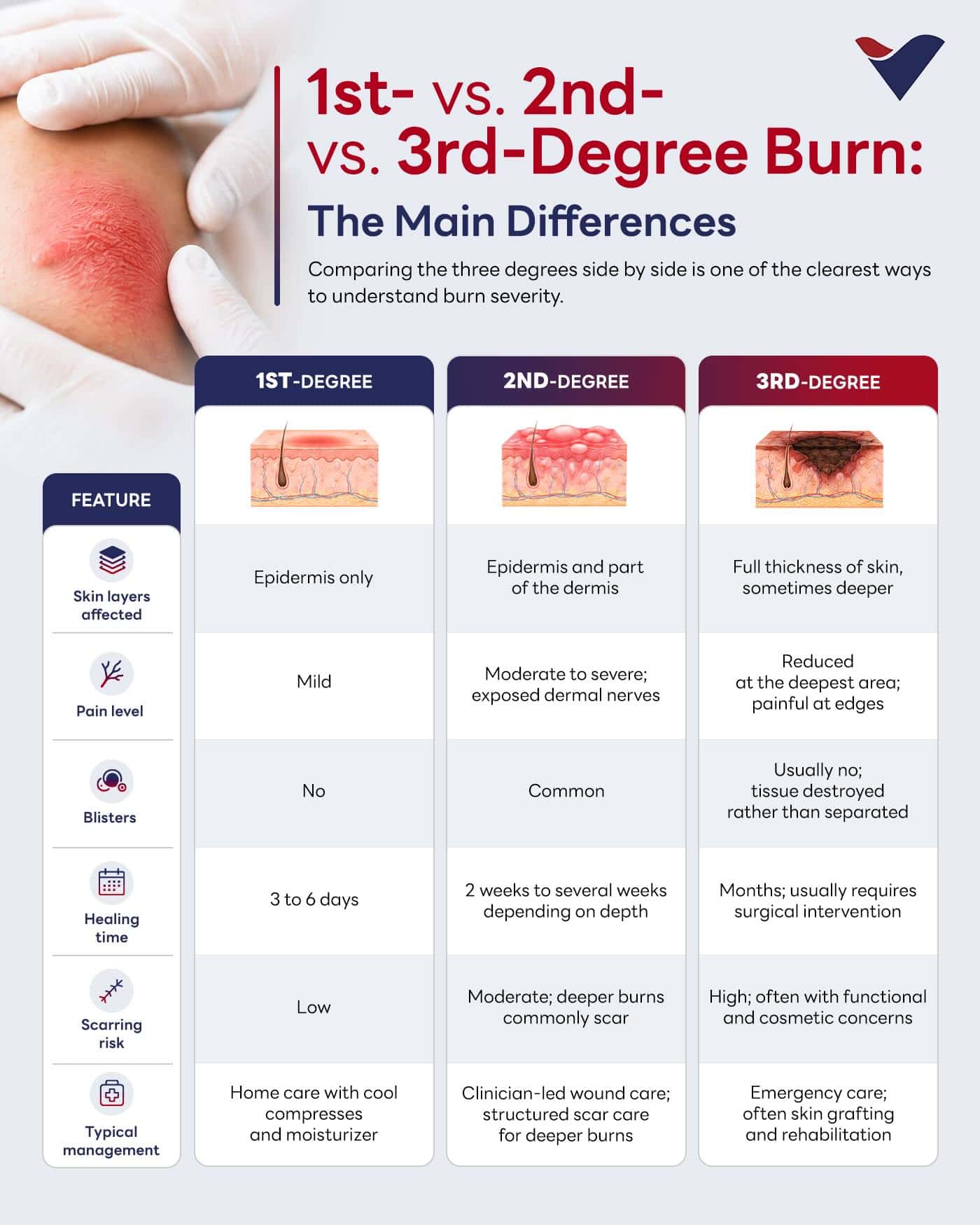
1st- vs. 2nd- vs. 3rd-Degree Burn: The Main Differences
Comparing the three degrees side by side is one of the clearest ways to understand burn severity.
| Feature | First-Degree (Superficial) | Second-Degree (Partial-Thickness) | Third-Degree (Full-Thickness) |
|---|---|---|---|
| Depth | Epidermis only | Into the dermis | Through epidermis + dermis |
| Appearance | Red, dry | Red/pink, moist, blistered | White, brown, black, leathery |
| Pain | Mild | Severe | Center may feel numb; edges hurt |
| Blisters | No | Yes | Not typically |
| Healing | ~3–6 days | ~2+ weeks | Requires surgery; long recovery |
| Scarring | Rare | Possible (esp. deep) | Very likely |
| Care | Home care | Home + medical review | Emergency / specialist |
Which Degree of Burn Is the Worst?
Third-degree (full-thickness) burns are the most severe in routine classification, because they destroy the entire thickness of the skin and can involve deeper tissue. Where a burn reaches muscle or bone, it may be classed as fourth-degree — the most dangerous of all and a medical emergency.
Burn First Aid: What to Do Right Away
For minor (first-degree and small second-degree) burns:
- Cool the burn under cool running water for 20 minutes — not ice.
- Remove tight items (rings, watches) before swelling starts.
- Cover loosely with a clean, non-stick dressing.
- Do not apply butter, oil, toothpaste, or ice.
- Take OTC pain relief as needed.
Call emergency services immediately if the burn is:
- Large (broadly, bigger than the person’s palm), deep, or full-thickness
- On the face, hands, feet, joints, or genitals
- Caused by electricity, chemicals, or fire in an enclosed space (inhalation risk)
- In a child, older adult, or person with diabetes or circulatory conditions
Treatment Options For Burn Scars
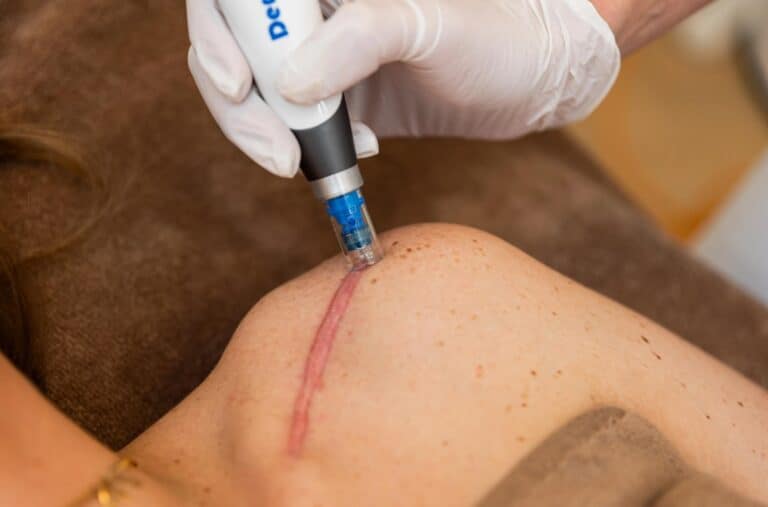
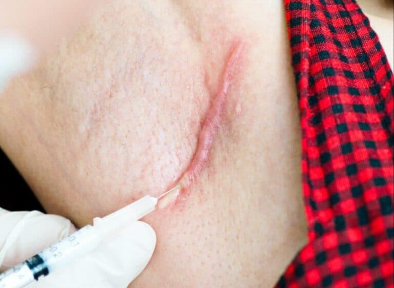
Burn scars can affect skin flexibility, restrict movement near joints, and produce ongoing itching or tightness. The depth of the original burn is the single biggest predictor of how the skin will recover, and several other factors, including wound care quality, infection, healing time, skin tone, and a personal or family history of keloid scarring, also influence the final result.
Begin your treatment for burn scars early and consistently for the best wound management. Several approaches may be combined depending on scar type, location, and stage of healing.
- Scar management therapies: Pressure therapy, silicone-based sheets or gels, regular moisturization, and ongoing sun protection to limit pigmentation changes.
- Wound care programs: Structured follow-up to monitor healing tissue and address concerns before they progress.
- Reconstructive and rehabilitation approaches: Appropriate for scars that restrict movement or affect function, particularly near joints.
- Supportive regenerative protocols: For suitable candidates, regenerative approaches under physician supervision may be considered as part of a staged scar care plan.
The earlier scar care begins, the more influence it can have on the final outcome. Even scars that have matured for months or years may still respond to physician-led intervention.
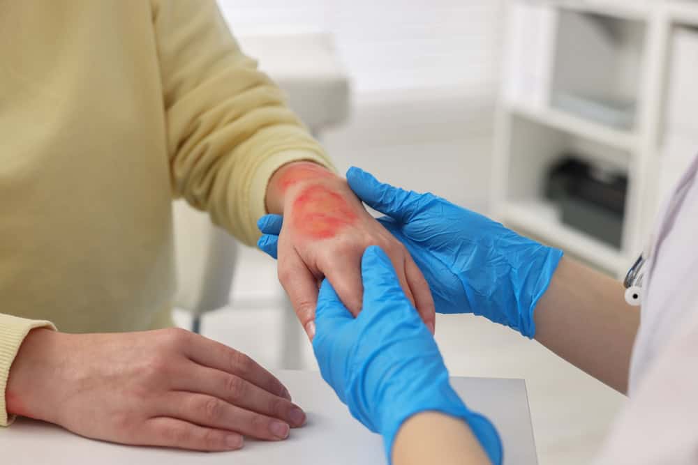
Why Early Wound Management Matters
The first few days of wound care often shape how skin heals over the following months — which is why timely clinical input matters even for burns that look manageable at first.
The case for early treatment
- Reduces complications: prompt cleaning, dressing, and assessment limit secondary damage, infection, and tissue loss — all of which drive scar formation.
- Supports healing: proper care shapes the recovery trajectory when the wound environment is most influential.
- May improve long-term outcomes for skin quality, joint function, and visible scarring.
Burns that always warrant professional evaluation: large burns; burns to the face, hands, feet, joints, or genitals; deep, electrical, or chemical burns; and any burn in a child, older adult, or person with diabetes or circulatory conditions.
Waiting to “see how it heals” can delay the care your skin needs. When in doubt, having a clinical team review the wound is the safer route.
The Vega Dermatology Approach
At Vega Dermatology & Wound Care, scar reduction is treated as a pathway from the earliest point of healing, not a single intervention after scars form:
- Physician-led wound assessment of burn depth, healing trajectory, and scar risk to guide an individualized plan.
- Scar care and tissue-remodeling programs with structured monitoring through scar maturation, plus pressure therapy and silicone options as appropriate.
- Regenerative protocols for suitable candidates, where supportive approaches such as VEGF and PDGF Ultra Enhanced Media may be considered under physician supervision to support tissue conditions during healing.
- Continuity of care — the same clinical team manages assessment, treatment, and follow-up, so scar decisions are made with full visibility of how the burn healed.
Early physician-led input is one of the most direct ways to support appropriate treatment for burn scars and protect long-term skin health. Book a consultation for a wound-care evaluation or scar assessment.

The Vega Derma Medical Team consists of board-certified dermatologists, specialized trichologists, and clinical researchers based in Bangkok, Thailand. Our team specializes in evidence-based regenerative dermatology, complex tissue repair, and advanced hair restoration (including precision FUE and FSE). Every protocol and article we publish is medically reviewed to ensure it meets international clinical standards, providing safe, science-backed guidance for our local and international patients.