

Introduction
People often use “stem cell” as a catch-all term, which creates confusion. A lab might take cells from fat, bone marrow, or umbilical cord tissue and label them as an MSC product, but proving they meet the standard is another matter. There is a real technical standard for what qualifies, and it was set nearly twenty years ago by a committee that wanted to bring clarity to the field.
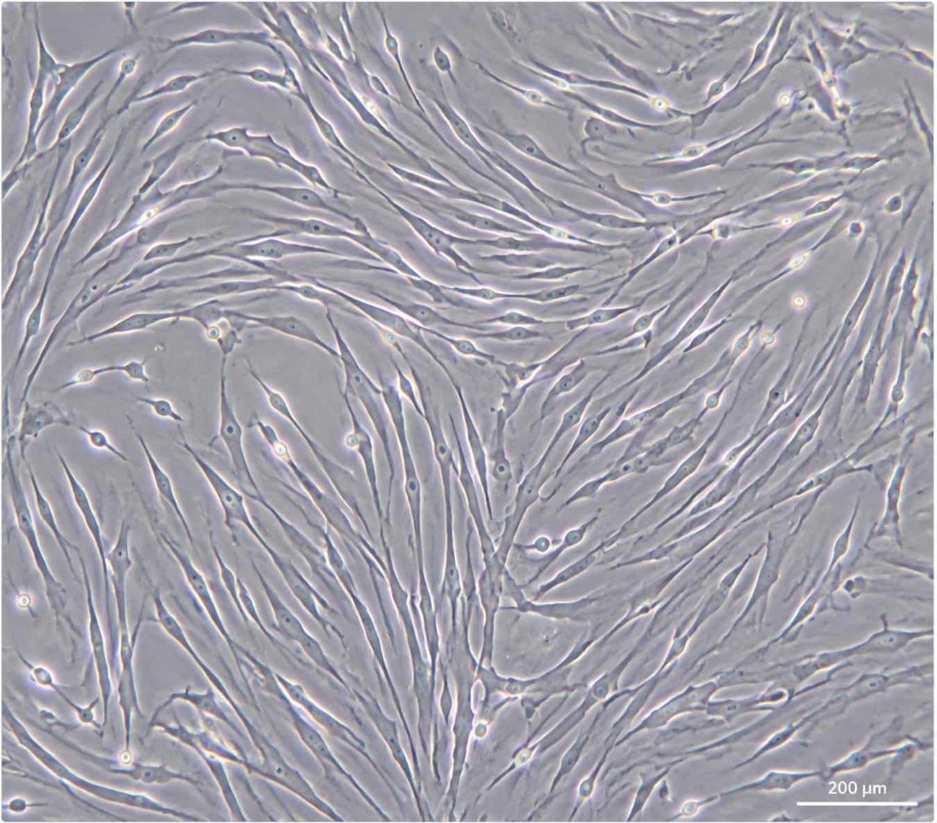
In 2006, the Mesenchymal and Tissue Stem Cell Committee of the International Society for Cellular Therapy (ISCT) published a short position paper that became the field’s main reference. The paper did not try to answer every scientific question about what a mesenchymal stromal cell is at the biological level. Instead, it set a basic standard: three conditions. If a cell population does not meet all three, it cannot be called an MSC. This standard helps separate labs that can support their claims from those that use the term too loosely. It is also why serious stem cell therapy providers in Bangkok, Thailand base their quality documentation on it.
Criterion One: Plastic Adherence
The first test seems almost too simple. Under standard culture conditions, the cells just need to stick to the plastic surface of a tissue culture flask.
This step is more important than it might seem. When bone marrow and cord tissue are first collected, they contain a mix of cells: hematopoietic cells, blood cells, debris, and the stromal cells that labs are actually looking for. Hematopoietic cells usually do not stick to plastic under standard culture conditions, but stromal cells do. This difference allows labs to wash away non-adherent cells after the first 24 to 48 hours, helping them focus on the cells they want. It is a basic filter, but it is the first one, and every MSC preparation must pass it before more advanced tests are used.
Adherence alone does not prove much, since many non-MSC cells, such as ordinary fibroblasts, also adhere to plastic. That is exactly why the ISCT included more tests.
Criterion Two: The Surface Marker Profile
When people talk about “the ISCT criteria,” they usually mean this standard. It actually involves two tests occurring simultaneously: positive selection and negative exclusion. Both are measured using flow cytometry.
Positive markers: At least 95% of cells must express the three surface antigens: CD73, CD90, and CD105. These markers are not random. CD73 is an ecto-5′-nucleotidase involved in adenosine signaling. CD90 (also called Thy-1) is a glycoprotein important for cell adhesion and signaling. CD105 (endoglin) acts as a TGF-β co-receptor. None of these markers is unique to MSCs, which is the main challenge. CD73 is also found on some epithelial and endothelial cells, and CD90 appears on certain neurons and fibroblasts. What matters is that all three are present together at high levels on the same cell, which helps identify MSCs more specifically.
Negative markers: At the same time, no more than 2% of the cells should show CD34, CD45, CD14, CD11b, CD79α, CD19, and HLA-DR. This part of the test helps make sure there is no contamination from other cell types that are often mistaken for MSCs. CD45 is a marker found on all leukocytes, so if it is present, hematopoietic cells have not been fully removed. CD34 marks hematopoietic progenitor and endothelial cells. CD14 and CD11b identify monocytes and macrophages, CD79α and CD19 identify B cells, and HLA-DR marks activated antigen-presenting cells. If a sample is positive for the first three markers but also has significant levels of any of these five, it is not a pure MSC preparation. Instead, it is a mix that includes MSC-like cells.
Taken together, this is the profile that labs look for when they use flow cytometry to check a batch before release: CD73+/CD90+/CD105+, CD34-/CD45-/CD14-/CD79a-/HLA-DR-. This is a specific and testable signature, not just a general description.
Criterion Three: Tri-Lineage Differentiation
The final criterion focuses on what the cell can actually do, not just how it looks. To be considered an MSC, the cell population mumust be able to turn into three different types of cells in the lab when given the appropriate conditions: osteoblasts (bone-forming cells), adipocytes (fat cells), and chondroblasts (cartilage-forming cells).
This process is how scientists prove that the cells are truly multipotent. In the lab, they test the cells with three different induction methods: osteogenic, adipogenic, and chondrogenic media. They then use specific stains to confirm the results: Alizarin Red shows calcium deposits in bone cultures, Oil Red O highlights fat droplets in adipogenic cultures, and Alcian Blue or Safranin O reveals proteoglycans in cartilage cultures. If the cells have the right surface markers but cannot show all three types of differentiation, they do not meet the ISCT definition. Surface markers indicate what the cells are, while trilineage differentiation indicates what they can do. The ISCT requires both.
Why This Framework Is the One That Matters
This is not just an academic detail. The ISCT criteria are important because without a common technical standard, “MSC” becomes a marketing term rather than a scientific one. That difference is where quality can vary the most between providers offering stem cell therapy in Bangkok, Thailand, or anywhere else.
Any clinic or lab that produces MSCs should be able to show flow cytometry data confirming the marker profile, along with documentation of tri-lineage differentiation testing for its cell bank. This is not just a nice extra, but the basic proof that what is being given is truly what it claims to be. If a provider cannot show this documentation, it does not always mean they are acting in bad faith, but it does mean they cannot be sure their product meets the internationally recognized definition of an MSC. It is important to ask about this directly before comparing anything else, such as protocol, dose, or price, because those details do not matter much if the cell identity has not been confirmed first.
There is also a related question that often comes up and deserves a full answer: what CD90 does, why it is one of the three positive markers rather than a negative one, and what it indicates about a cell population on its own. This is a complex topic that should be covered in its own article. Here, I am just providing the framework for that discussion.
The Takeaway
There are three tests, done one after another: the cells must stick to plastic, show the right surface markers, and be able to turn into three different tissue types. That is the basic requirement. This does not describe what MSCs can do or how they are used in medicine. Instead, it is just the minimum standard for designating a group of cells as MSCs, and it should serve as the basis for any other claims a lab or clinic makes about them.
Frequently Asked Questions
Does this hurt, and is there downtime?
The injection step can feel mildly uncomfortable, but local anesthetic keeps that manageable for most patients. Downtime is minimal most people are back to normal activities the same day.
How many sessions does it take to see results?
Generally two to three sessions, spaced three to four months apart, with the clearest results typically showing up somewhere between six and twelve months after starting.
Is stem cell therapy actually safe?
When it’s performed by an experienced physician under proper medical standards, the overall risk is low especially when the cells or extract come from the patient’s own body rather than a donor source.
The Bottom Line
Stem cell therapy for hair loss represents a real shift in how hair restoration works, going straight after the stem cells at the root of the problem instead of just boosting blood flow or reinforcing hair that’s already growing. For patients with dormant follicles that haven’t fully atrophied, it opens up an option that genuinely wasn’t available a few years ago.
A consultation with a specialized team at a clinic like Vega Derma for a real candidacy assessment and a treatment plan built around your specific follicle condition is the right first step toward a result that’s both lasting and safe.

The Vega Derma Medical Team consists of board-certified dermatologists, specialized trichologists, and clinical researchers based in Bangkok, Thailand. Our team specializes in evidence-based regenerative dermatology, complex tissue repair, and advanced hair restoration (including precision FUE and FSE). Every protocol and article we publish is medically reviewed to ensure it meets international clinical standards, providing safe, science-backed guidance for our local and international patients.