Case Details
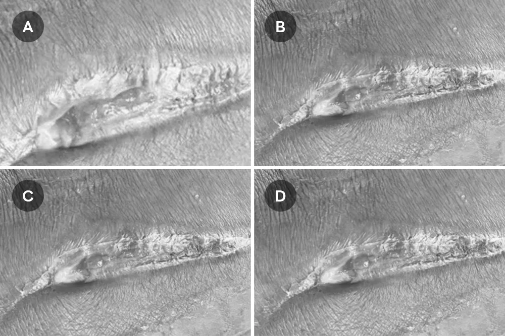
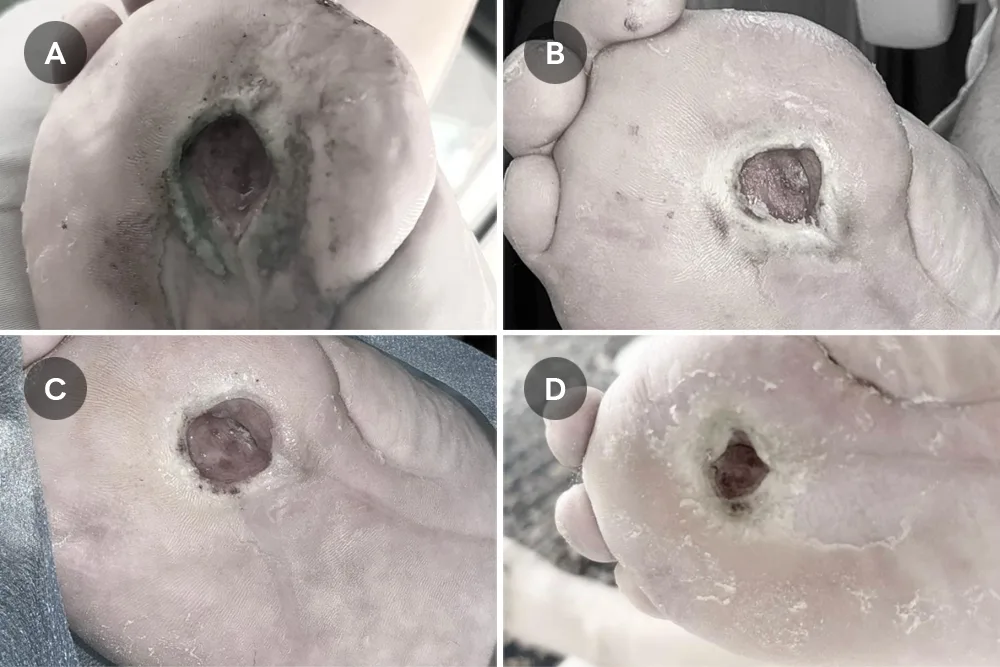
Serial photographs of a chronic diabetic wound at the right plantar foot during combined UC-MSC-based treatment and wound care.
(A) Pre-treatment appearance showing a chronic plantar ulcer with prominent surrounding necrotic tissue.
(B) Baseline at treatment initiation (23 January), with wound size approximately 12 mm after debridement.
(C) Day 10 follow-up (2 February), showing reduction in wound size to approximately 8
mm, less necrotic tissue, and improved wound bed appearance.
(D) Follow-up image taken on 19 February, demonstrating continued improvement with a more consolidated granulation bed, reduced periwound maceration, and clearer early signs of wound contraction.