Case Details
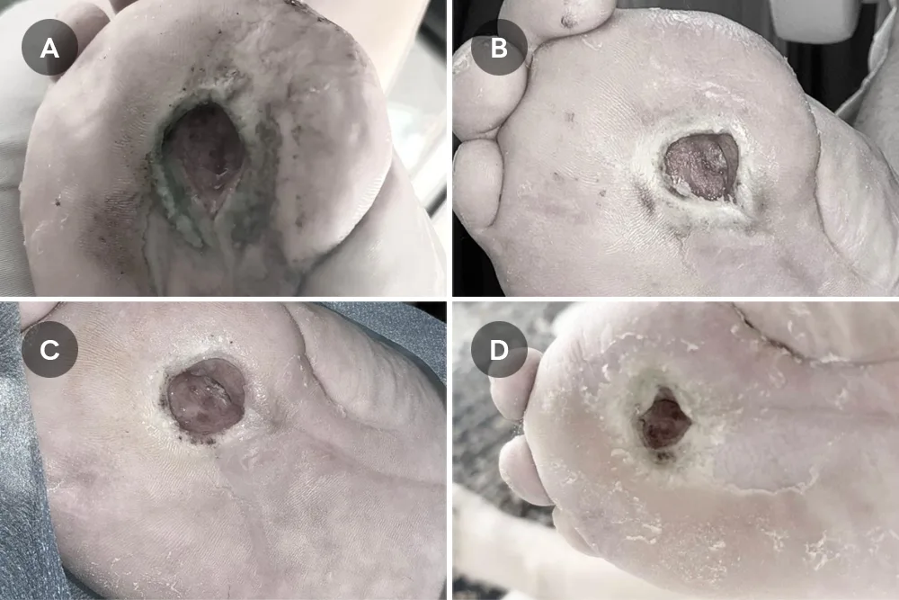
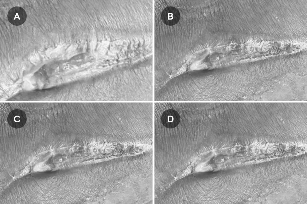
Figure 1. Serial clinical photographs of a postoperative dehisced wound showing progressive healing during UC-MSC–assisted regenerative therapy and wound care.
(A) Day 1: Initial presentation demonstrating a dehisced linear postoperative wound with exposed granulation tissue and substantial adherent yellow slough along the wound bed. Surrounding skin shows erythema and early maceration.
(B) Day 5: Early follow-up shows persistent wound dehiscence with mixed slough and fibrin coverage, though the wound margins appear more clearly defined with reduced surrounding inflammation compared with Day 1.
(C) Day 10: Intermediate follow-up demonstrating gradual improvement, featuring a reduced slough burden, healthier emerging granulation tissue, and partial reduction in wound depth.
(D) Day 14: Subsequent follow-up revealing continued healing progression with further consolidation of granulation tissue, decreased exudate and slough, and early signs of wound contraction along the wound edges.